Selasa, 31 Agustus 2010
EFEK BIOLOGI PADA PEMERIKSAAN MRI
Medan magnet, dalam ilmu Fisika, adalah suatu medan yang dibentuk dengan menggerakan muatan listrik (arus listrik) yang menyebabkan munculnya gaya di muatan listrik yang bergerak lainnya. (Putaran mekanika kuantum dari satu partikel membentuk medan magnet dan putaran itu dipengaruhi oleh dirinya sendiri seperti arus listrik; inilah yang menyebabkan medan magnet dari ferromagnet "permanen"). Sebuah medan magnet adalah medan vektor: yaitu berhubungan dengan setiap titik dalam ruang vektor yang dapat berubah menurut waktu. Arah dari medan ini adalah seimbang dengan arah jarum kompas yang diletakkan di dalam medan tersebut.
EFEK BIOLOGI :
- medan magnet bumi tidak memberi dampak pada kehidupan makhluk kecil
Mis: burung migrasi terkena 6 gauss.
- penelitian pd monyet dengan 10 T magnet tidak ada efek yg membahayakan.
- hasil penelitian penggunaan magnet < 2 T tidak ada efek pd pertumbuhan atau morfologi sel
SEBELUM KITA BICARA LEBIH LANJUT TENTANG DAMPAK MRI KITA BELAJAR DULU BARANG2 APA SAJA YANG TIDAK BOLEH MASUK MRI
Implantasi pd pasien dapat menimbulkan serius efek spt : torque (berputar), heating, artifact pd gbr MRI.
1.Jam analog
2.Tape recorder
3.Credit card
4.Calculators
5.Perhiasan
6.Wigs
7.Jepit rambut
8.Gigi palsu dll
9.Pasien dengan pacemaker
10.Pasien dengan klip aneurisme
Bahan ferromagnetik dpt berbahaya
1.Bahan feromagnetik semua tertarik oleh magnet. Klip kertas atau hair pins – bila tertarik ke magnet (1,5 T) kecepatan hingga 60 km/jam.
2.Alat bantu lain : hemostats, gunting, klem yg terbuat dari stainless steel juga tertarik magnet
3.Tangki oksigen juga dpt ditarik oleh magnet
4.Petugas lain spt perawat, cleaning service, pemadam kebakaran dll harus diberi penjelasan tentang bahaya magnet tsb.
5.Di depan ruangan, harus ditempelkan tanda
6.Masyarakat umum harus berada di area 5-15 G
7.Screening dengan metal detector bila ada.
EFEK JIKA MEMAKAI BARANG-BARANG SEPERTI DIATAS
Aneurisme klips : kontra indikasi MRI .Hanya akan diperiksa dengan MRI bila klips tsb pasti non ferromagnetic.
- Protehese : pd katup jantung : dr penelitian diketahui bahwa 25 dr 29 prothesis terpengaruh oleh magnet, akan tetapi pergerakannya masih lebih kecil dibanding dgn akibat pergerakan jantung. Model Pre-6000 kontra indikasi MRI.
continued….
Sabtu, 28 Agustus 2010
MEDIA KONTRAS MRI
•Contras media MRI peningkatan pencitraan yg diperoleh dari absorpsi gelombang radio oleh inti atom
•Contras media MRI adalah bahan paramagnetic atau superparamagnetic yg meningkatkan citra resonansi magnetic dengan mempengaruhi waktu relaksasi dari inti
•Media kontras paramagnetic meliputi kompleks gadolinium seperti gadodiamid, gadopentetic acid dan gadoteridol dan kompleks mangan
•Superparamagnetic meliputi Kompels iron, ferrocarbitrans dan ferromoksil
Indikasi pemberian kontras pada MRI :
1.MRI Brain
Indikasi :
•Neoplasia
•Infection
Infarction
2.MRI Spine
Indikasi :
•Degenerative disc disease
•Inflamatory lesions
•Neoplasia
•Vascular disease
•Demyelination
•Conginetal lesions
3.MRI Whole Body
Indikasi :
•Bronchogenic carcinoma
•Acute myocardial infarction
•Breast imaging
•Liver and spleen
•Bowel
•Kidney
•Adrenal gland
•Pelvis
•Musculoskeletal system
KONTRA INDIKASI Untuk Pemeriksaan MRI
•Mutlak :
–kehamilan dan menyusui
–Gagal ginjal
•Relatif :
–Anemia hemolitika
–Riwayat reaksi alergi dengan bahan iodida
TYPE MEDIA KONTRAS MRI
•Intravenous
–Extracelluler distribution
•Gd DTPA (Gadopentetate dimeglumin)
•Gd HP-DOGA (Gadoteridol)
•Gd DTPA-BMA (Gadodiamide)
•Gd DCTA
–Hepatobillary
–Particulate
•Oral
–Metal chelat
•GD DTPA
–Magnetic Iron Oxide Particles
•Oral Magnetic Particles
•AMI-121
7T MRI* Explore the Future of MRI. Today.
Siemens is the world leader in MRI technology and applications development. With more and more applications calling for higher resolution images, ultra-high-field MRI technology has become compulsory for leading institutions. 3T MRI has become clinical reality in order to answer complicated questions and to leverage the transfer of research methods into useful clinical applications. 7T MRI* now emerges as the equipment of the elite, the new research and development frontier, the system of the pioneers.
7T MRI - a great research instrument
7T MRI provides the potential for microscopic spatial resolution visualizing anatomy previously unseen. In addition, it enables the observation and analysis of tissue metabolism and function. 7T MRI is a great instrument for the research and development of molecular imaging methods, promising a whole new world of applications. High-level MR hardware and software expertise on site are prerequisites for the successful operation of such a system. 7T MRI systems are investigational devices and are not available for clinical use (i.e. it can not be used for clinical diagnosis but only for clinical research purposes).
7T MRI brings certain types of MRI research, such as ultra-high resolution human fMRI (0.1mm resolution, which reach slowly the level of resolution provided by histology), out of the animal realm for the first time. It will eventually become possible to study neuronal function at the sub-millimeter scale. Potential clinical applications include neurodegenerative diseases (Alzheimer, etc.). The focus is currently set on brain imaging but other applications in the rest of the body are not excluded.
The Siemens 7T MRI program includes today:
- 7T Head Scanner: 90 cm bore size magnet. Actively shielded 2.20 m long magnet with 68 cm bore diameter, enables siting of 7T in a conventional 3T foot print.
- 7T Whole-Body Scanner: Unshielded 90 cm bore size magnet witch is 3.40 m long.
Siemens 7T MRI projects
Siemens Healthcare is the leading vendor in 7T ultra-high-field MRI. More than half of the 30 installed systems worldwide are from Siemens. The success of the Siemens ultra-high-field program is explained by the technological leadership provided by Siemens and in particular: the 32 independent RF channels of the Tim technology, the strong, fast and reliable gradients as well as the most advanced application methods. The success is also explained by the strong collaboration between the research institutes hosting the 7T systems and Siemens, which is not only necessary but a prerequisite for full exploitation of the capabilities of such a system.
Siemens collaboration with the CEA
The Commissariat à l’Energie Atomique (CEA) with headquarters in Paris and Siemens Healthcare agreed to expand and intensify their joint research activities in the area of innovative imaging and therapy. This included the area of ultra-high-field MRI, the development of 11.7 T systems for human applications as well as 17 Tesla systems for small animal research should lead to breakthroughs in the areas of diagnosis and therapy for neurological diseases, such as Alzheimer’s or Parkinson’s disease. At present, the upper limit for human applications is 7 Tesla.
"Siemens is the ideal partner for us to transfer our comprehensive know-how and long-term experience in the area of medical imaging, diagnostics and therapy from research to clinical routine," says Alain Bugat, Administrateur Général of CEA. “There is no doubt in my mind that as a team we are able to move medicine a giant step forward.“
Jumat, 27 Agustus 2010
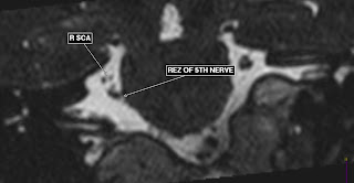
PERANAN MRI PADA KASUS TRIGEMINAL NEURALGIA
Pernah merasa nyeri Gusi,Gigi dan Wajah ?
Atau Nyeri seperti tersengat listrik dan di tusuk jarum ?
Senin, 23 Agustus 2010
Komunitas Hemifacial Spasm Indonesia
Nah untuk para penderita HFS tidak perlu kuatir karena penyakit ini sudah bukan menjadi momok lagi Anda bisa berkonsultasi dan berkomunikasi dengan para penderita HFS Komunitas Hemifacial Spasm
Semoga informasi ini bermanfaat
Minggu, 22 Agustus 2010
New Info Saatnya Belajar MRI
Para pecinta MRI dan penggemar blog saya terima kasih atas atensinya sehingga blog ini banyak memberikan manfaat bagi semua orang.
Karena materi yang saya tulis sudah terlalu banyak maka untuk mencari informasi tentang MRI Anda bisa ikuti langkah saya :
Step 1. Buka http://belajar-mri.blogspot.com/
Step 2. Pikirkan informasi apa yang ingin Anda ketahui ?Jika sudah ketemu
Step 3.Lihat ada gedget Cari Informasi disini Anda ketikkan Materi yang Anda mau lihat
contoh Ketik Spondylosis maka akan ada materi saya di sisi kiri.Atau Anda bisa cari di link yang lain.
Nyeri Kepala ternyata Schwannoma
Schwannoma
Ok hari ini kita belajar tentang Schwanoma apa itu dan bagaimana Pemeriksaan MRI nya.
Schwannoma adalah sel jinak yang dimulai dalam sel schwan yang menghasilkan myelin yang melindungi acustic (wikipedia).Biasanya ditemukan dicerebropontine.
Penyebabnya belum diketahui meskipun ada literatur yang bilang adalah kelainan genetik (wikipedia)neurofibromatosis merupakan faktor resiko.
berbicara nerofibromatosis kita juga harus tahu apakah itu ? betul gak ?
Neurofibroma adalah benjolan seperti daging yang lembut, yang berasal dari jaringan saraf.
Neurofibroma merupakan pertumbuhan dari sel Schwann (penghasil selubung saraf atau mielin) dan sel lainnya yang mengelilingi dan menyokong saraf-saraf tepi (saraf perifer, saraf yang berada diluar otak dan medula spinalis).
Pertumbuhan ini biasanya mulai muncul setelah masa pubertas dan bisa dirasakan dibawah kulit sebagai benjolan kecil.
Neurofibromatosis bisa mengenai setiap saraf tubuh tetapi sering tumbuh di akar saraf spinalis. Neurofibroma menekan saraf tepi sehingga mengganggu fungsinya yang normal.
Neurofibroma yang mengenai saraf-saraf di kepala bisa menyebabkan kebutaan, pusing, tuli dan gangguan koordinasi.
Semakin banyak neurofibroma yang tumbuh, maka semakin kompleks kelainan saraf yang ditimbulkannya
saya menggunakan sequence rutin untuk MRI brain tetapi saya tambahi dengan teknik T2 DRIVE HR ini merupakan teknik 3D T2, teknik ini sangat bagus sekali untuk mengetahui saraf2 di akustikus.Saya akan jelaskan mengenai teknik T2DRIVE HR=T2 Weighted 3D turbo spin echo sequence INFO:TR is relatively short to reduce scan time.heavy T2-weighted can still be obtained by using DRIVE
Gb.1 T1 W pre contras Gb.2 T1 W post contrast

Gb.1 T1 W pre contras Gb.2 T1 W post contrast
Jika dilihat gambar di atas terlihat jelas area enhancment yang menandakan adanya massa.
Jumat, 13 Agustus 2010
metastases in mylum techniques found in the MR total spine
In case study women age patient 73 th come to the doctor with clinical neurosurgery cervical spondylosis and lumbalis going to the Husada Utama Hospital to investigate MR Whole spine ( otal Spine )
Discreening patient by Nurse ask whether there are MRI in pace maker what is not? Use carotid clip or not to whether a history of surgery or chemotherapi if ever done or no ?
Radiografer to explain how to long this examination and inspection procedure
1.Patient confertable in supine postition
2.Normal breathing
3.During examination must not be moved
By using the technique that is very sequence Pasting direcly from cervical untul sacrum Sequence once took approximately 4 minute T2 Weighted. T1 weighted. Stir Long TE (ie longer 5 Minute) and axial slice T2Weighted and T1 weighted taken at the vertebral bone join
THis examination is usually in use for the case :
1.Metastase
2.Spondylosis cervicalis
3.Spomdylosis Lumbalis and
4.Scoliosis
There are some case with whole spine examination found the process of metastase in myelum.The examination very helpful for screning
by.ferry Indriasmoko


